Endometriosis Diagnosis: The Modern Pathway
Diagnosing endometriosis is no longer a single test. It is a structured pathway that combines medical history, symptom pattern, pelvic examination when appropriate, expert-protocol imaging, and — when needed — laparoscopy with tissue biopsy for histologic confirmation.
Why endometriosis diagnosis is difficult
Endometriosis lesions can be small, deep, or located in areas not seen on a standard pelvic ultrasound. Symptoms overlap with more common conditions such as IBS, bladder pain syndrome, and musculoskeletal pain, and there is currently no validated blood test for the disease. As a result, patients experience an average diagnostic delay of 6 to 10 years in most published data. A structured, specialist-led diagnostic pathway shortens that delay.
Step 1 — Symptom review and medical history
The first step is a structured clinical interview covering:
- Menstrual pain pattern and progression
- Non-menstrual pelvic pain, deep dyspareunia, and cyclic bowel or bladder symptoms
- Fertility history and goals
- Prior treatments — hormonal therapy, prior surgery, prior imaging
- Family history of endometriosis or chronic pelvic pain
- Impact on quality of life, school, work, and mental health
A structured history often identifies patterns that suggest deep infiltrating disease, bowel or ureteral involvement, or coexisting adenomyosis — all of which shape the imaging plan.
Step 2 — Clinical examination
When appropriate and acceptable to the patient, a pelvic examination may identify tenderness of the posterior fornix, nodules in the recto- vaginal septum, uterosacral thickening, a fixed retroverted uterus, or pelvic floor dysfunction. Normal examination does not rule out disease. Examinations should always be trauma-informed and patient-consented.
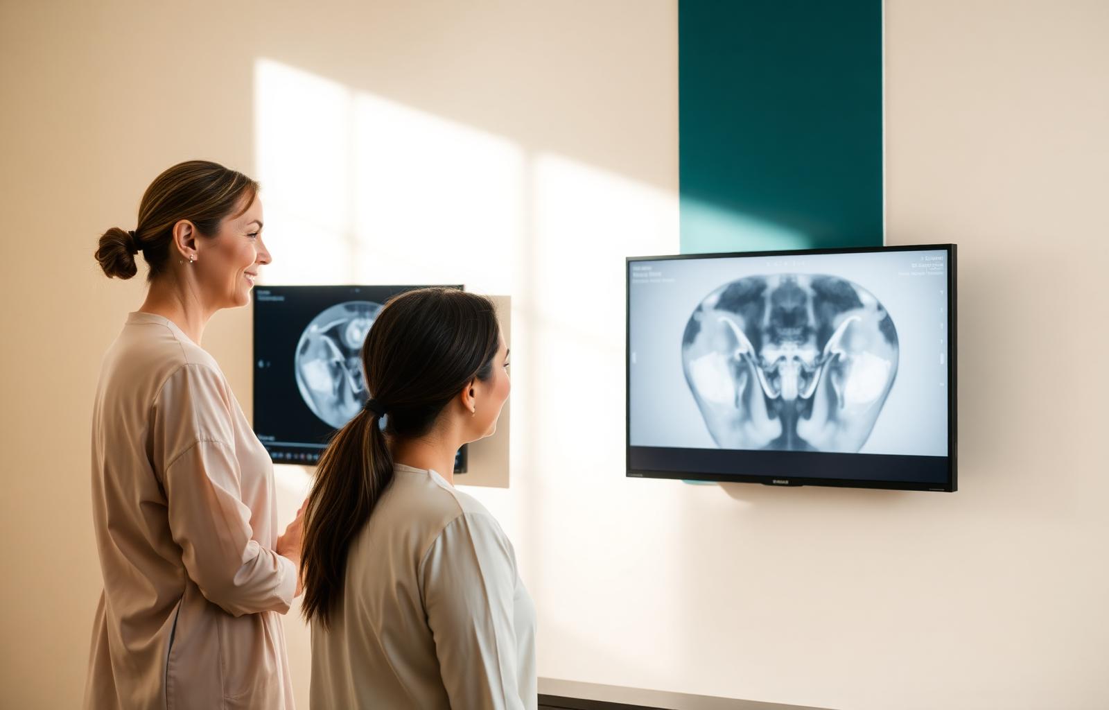
Step 3 — Expert-protocol ultrasound
A dedicated endometriosis ultrasound — sometimes called a mapping or deep-endometriosis ultrasound — is performed by trained sonographers using a defined protocol to evaluate:
- Uterus (including signs of adenomyosis)
- Ovaries (including endometriomas)
- Posterior compartment (uterosacral ligaments, recto-vaginal septum, bowel)
- Anterior compartment (bladder)
- Ovarian and uterine mobility (a marker of adhesions)
A standard pelvic ultrasound is not the same test. A normal standard ultrasound does not rule out endometriosis.
Step 4 — MRI with endometriosis protocol
MRI performed with an endometriosis-specific protocol and read by trained radiologists is used to:
- Map deep infiltrating endometriosis
- Evaluate bowel, bladder, ureteral, and pelvic sidewall involvement
- Assess adenomyosis
- Help plan a multidisciplinary surgery when needed
Imaging accuracy depends heavily on protocol, equipment, and reader expertise. Review by an experienced endometriosis center is often what changes the diagnosis.
Step 5 — Laparoscopy with histologic confirmation
Laparoscopy remains important when:
- Symptoms and imaging do not match
- Imaging cannot be performed with an expert protocol
- Tissue confirmation is needed
- Surgical treatment is planned for pain, fertility, or organ involvement
When laparoscopy is performed, lesions should be excised whenever possible and sent for pathology. Diagnostic-only laparoscopy without biopsy or treatment is discouraged in modern practice.
What to bring to a diagnostic consultation
- A timeline of symptoms and prior treatments
- Copies of prior imaging (CDs and reports)
- Operative and pathology reports from prior surgeries
- List of current medications and hormonal treatments
- Fertility goals and any prior fertility evaluation
What a good diagnosis looks like
A good diagnostic workup ends with a clear map of what is happening, which structures may be involved, what still needs clarification, and what treatment options exist — including watchful waiting, medical management, fertility planning, and surgery. It is a plan, not just a label.
Continue with MRI & Ultrasound Mapping, Endomapping & Surgical Planning, or — if surgery is being considered — Excision vs Ablation and how to choose a surgeon.
Related pages
Speak with an endometriosis advisor
Share your symptoms, prior treatment, and goals. An advisor will help you understand your options and connect you with the appropriate specialists.
Frequently asked questions
›How is endometriosis diagnosed today?
Diagnosis combines symptom review, medical history, pelvic examination when appropriate, expert-protocol ultrasound and MRI, and — when imaging is inconclusive or when treatment is indicated — laparoscopy with tissue biopsy for histologic confirmation.
›Can a standard pelvic ultrasound rule out endometriosis?
No. A standard pelvic ultrasound may appear normal even in patients with significant disease. Deep infiltrating endometriosis and small peritoneal lesions frequently require a dedicated endometriosis-mapping ultrasound protocol or MRI with expert interpretation.
›What is an endometriosis-mapping MRI?
It is an MRI performed with a specialized protocol and read by radiologists trained in endometriosis. It evaluates the uterus, ovaries, pelvic peritoneum, bowel, bladder, ureters, pelvic sidewall, and nerve regions to plan surgery.
›Is a blood test available for endometriosis?
There is currently no validated blood test that reliably diagnoses endometriosis. Research is ongoing. Diagnosis remains clinical, radiologic, and — when needed — surgical with histologic confirmation.
›When is laparoscopy still recommended?
Laparoscopy is considered when symptoms and imaging do not match, when tissue confirmation is needed, when surgery is planned for treatment, or when infertility evaluation requires direct visualization of the pelvis.
›Does a negative laparoscopy rule out endometriosis?
Not always. Lesions can be subtle, deep, or missed by less-experienced surgeons. If symptoms persist after a negative laparoscopy, a second opinion from a specialist center may be considered.
›Do I need to stop hormonal therapy before imaging?
This depends on the imaging protocol and the clinical question. Some centers prefer specific menstrual cycle timing; others do not. Follow the instructions of the imaging center and referring clinician.
Medical review notice
This page was written for patient education and reviewed for medical accuracy by a member of the EndoHelp Medical Review Board.
- Reviewed by
- Dr. Ramiro Cabrera Carranco, MD
- Specialty
- Medical Reviewer — Deep Endometriosis, Gynecologic Endoscopy & Reproductive Surgery
- Content reviewed
- Endometriosis diagnosis, excision surgery, patient navigation.
- Last reviewed
- January 2026
Selected sources
Medical review policy · Editorial policy · References & sources · Network transparency
This content is educational and is not a substitute for professional medical advice, diagnosis, or treatment. Always consult a qualified healthcare professional regarding your individual condition.